
Glaucoma/ Kala Motia
Glaucoma is a blinding disease of the eye. The symptoms are akin to Cataract (Safed Motia). It affects mostly in elderly population, though can occur in other ages even at birth (buphthalmos). It can occur in patients with Cataract and some other eye diseases.
It causes progressive loss of peripheral field of vision without patient realising it. Early diagnosis and simple treatment with eye drops helps to arrest further loss of peripheral vision. There are three significant tests to confirm the diagnosis.
1) Intra- ocular pressure (IOP): shows increased fluctuation and levels higher than normal range (15- 21 mm Hg
2) Field of vision: gets constricted progressively.

3) A depression is seen in the Optic Nerve Disc by fundus examination (Glaucomatous cupping)

The visual loss due to glaucoma is irreversible; hence early detection is the key to prevent damage to eye sight.
Glaucoma tends to have a genetic predisposition hence if there is a positive family history of glaucoma, routine screening must be done of family members to detect early disease.
There are three clinical entities of Glaucoma depending upon the structure of Angle of the Anterior Chamber.
- Open Angle Glaucoma (Primary- POAG)
- Closed Angle Glaucoma (Primary- CAG)
- Mixed Glaucoma (Features of both types as above)
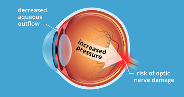
The Intra Ocular Pressure (IOP) : the eye maintains a pressure called Intra Ocular like the body maintains blood pressure in the blood vessels. Ciliary body inside the eye secretes nourishing fluid (Aqueous Humor) to provide nutrition to the crystalline lend and cornea the transparent lenses of the eye. These lenses do not have any direct blood supply. The aqueous humor after providing nourishment to the lenses find micro channels for their exit in the angle of the anterior chamber (trabecular meshwork). Fluctuation between secretion and drainage cause variation in the Intra Ocular Pressure. The normal range of variation in between 3-5 mm Hg. Fluctuation and high intra ocular pressure is responsible for damage to the periphery retina which causes loss of field of vision. The second possible cause of loss of peripheral vision is not related to the IOP, but is caused independently by a reduced blood supply to the peripheral retina. Thus there is clinical condition called as Normo-tensive Glaucoma.
This is the most common form of glaucoma in India. It occurs as a result of aging, the drainage angle of the eye becomes less efficient with time, and pressure within the eye gradually increases.
If the increase in pressure results in optic nerve damage, it is known as chronic open- angle glaucoma. Over 80% (?) of adult glaucoma patients have this type of glaucoma.
Chronic open-angle glaucoma damages vision so gradually and painlessly that one is not aware of trouble until the optic nerve has been extensively compromised.


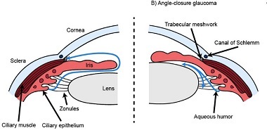
The drainage angle of the eye is examined by a test known as Gonioscopy. The drainage area (trabecular meshwork) can be obstructed by reduced entry, cellular debries to reduce progressively the aqueous humor resulting in chronic closed angle glaucoma. Total blockage of the entry by the root of the iris results in acute painful state due to sudden increase of the Intra Ocular Pressure (Acute Angle Closure Glaucoma – AACG) ( Fig 1 & 2).
Common Symptoms in Chronic Angle Closure Glaucoma are Rainbow color haloes around lights, eye aches and Blurred vision; while in in Acute Angle Closure Glaucoma the symptoms are Pain in the eyes, sometimes severe with Headache & Nausea and vomiting.
If you have any of these symptoms, call your ophthalmologist immediately. This is an emergency situation for the eye and requires urgent treatment. Acute angle closure glaucoma is more common in Asian people than in people of European descent; it is rare in people of African descent.

In some patients glaucoma has features of both the chronic open angle type and the acute angle closure type. This may be called chronic angle closure glaucoma or mixed mechanism glaucoma. It occurs more frequently in people of African and Asian descent.
Regular eye examinations by your ophthalmologist are the best way to detect early glaucoma.
During a complete and painless examination, you ophthalmologist will:
- Measure your intraocular pressure (tonometry): by Air Puff Method (Non-Contact) & Applanation Tonometry done on Slit Lamp Biomicroscope.
- Inspect the drainage angle of your eye (gonioscopy)
- Evaluate any optic nerve damage (ophthalmoscopy)
- Test the visual field of each eye (perimetry)
- OCT analysis of the Optic Nerve head & Retinal Nerve Fibre layer
- Measures the retinal nerve fiber layer thickness and Optic Nerve Cupping by equipment called OCT (Optical Coherence Tomography)
- Pattern ERG and Contrast Sensitivity for early diagnosis of Glaucomatous damage occurs in the ganglion cell layer of the retina. This change can occur few years before field defects typical of Glaucoma development
- Ocular Response Analyser- Further accurately evaluates Intra Ocular Pressure (IOP) , correcting for the dynamics of the eyeball ‘wall’ and IOP is read as IOPcc/IOPg.
Some of these tests may not be necessary for every person. Alternatively, you may need to repeat these tests on a regular basis (Annual), to monitor of the progress of the disease and review of the treatment.
As a rule, damage caused by glaucoma cannot be reversed. Eye drops, oral tablets, laser and surgical operations are used to check the progress of the disease.
With any type of glaucoma, periodic examinations are very important to prevent vision loss. Because glaucoma can worsen without ones’ being aware of it, Regular follow up is essential.
Glaucoma is usually controlled with eye drops taken several times a day, sometimes in combinations with oral tablets. These medications decrease eye pressure, either by reducing the production of aqueous fluids within the eye or by improving the out flow through the drainage channels.
For these medications to work, one must take them regularly without break. It is also important to tell physician about the eye medications you are using.
Glaucoma medications can have side effects. You should notify your ophthalmologist immediately if you think you may be experiencing side effects.
Some eye drops may not suit and cause: A stinging sensation, Red eyes, Changes in pulse and heartbeat, Changes in energy level, Changes in breathing (especially with asthma or emphysema), Headaches and Blurred vision.
Oral medicines may not suit some individual and causes: Tingling of fingers and toes, Drowsiness and Breathlessness and other systemic symptoms.
Laser surgery treatments may be effective for different types of glaucoma. The laser is usually used in one of two ways.
In open-angle glaucoma, the drain itself is treated. The laser is used to modify the drain (trabeculosplasty) to help control eye pressure.
In angle-closure glaucoma, the lasers create a hole in the iris (iridotomy) to create a by-pass a posterior to the anterior chamber within the eye to improve the flow of aqueous fluid.
When operative surgery is needed to control glaucoma, your ophthalmologist uses miniature instruments to create new drainage channel (Trabeculectomy) for the aqueous fluid to leave the eye. The new channel helps to lower the pressure.
Though serious complications of modern glaucoma surgery are rare, however, they can occur, as with any surgery. It is indicated if the IOP is not coming under control by medical treatment or if the compliance of the medical treatment is poor. Surgery is recommended if your ophthalmologist feels that it is safer to operate than to allow optic nerve damage to continue.
Some complicated cases of advanced glaucoma patients require special valve surgery to control of the eye pressure.
Some other indication for surgery are Cryotherapy, Glaucoma Valve implantation.
Don’t get dejected when diagnosed as Glaucoma Suspect. It is to protect you against a serious blinding disease. Rather feel satisfied to get it ruled out or treat.
If your parents are known case of Glaucoma, do tell the doctor to rule out Glaucoma by Special tests.
If you re diabetic and high myopia, do tell the doctor to rule out Glaucoma by Special tests.