Know Your Eye
Click on the image to see description.
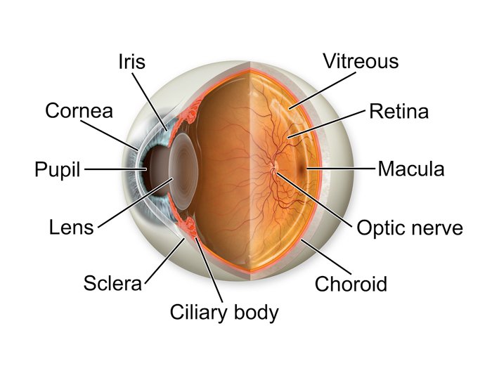
Pupil
The pupil is the opening in the center of the iris. The function of the pupil is to allow light to enter the eye so it can be focused on the retina to begin the process of sight. Typically, the pupils appear perfectly round, equal in size and black in color. The black color is because light that passes through the pupil is absorbed by the retina and is not reflected back (in normal lighting).
Iris
Iris is a thin, annular structure in the eye, responsible for controlling the diameter and size of the pupil, thus the amount of light reaching the retina. Eye color is defined by that of the iris.
Cornea
Cornea is the transparent part of the eye that covers the front portion of the eye. It covers the pupil, iris, and anterior chamber (the fluid-filled inside of the eye). The cornea's main function is to refract, or bend, light.
Sclera
The sclera is the white part of the eye that surrounds the cornea. In fact, the sclera forms more than 80 percent of the surface area of the eyeball, extending from the cornea all the way to the optic nerve, which exits the back of the eye. Only a small portion of the anterior sclera is visible.
Lens
The lens allows the eye to focus on objects at varying distances. It is located behind the iris and in front of the vitreous body.
Vitreous
The vitreous humour (also known simply as the vitreous) is a clear, colourless fluid that fills the space between the lens and the retina of your eye. 99% of it consists of water and the rest is a mixture of collagen, proteins, salts and sugars. Despite the water-to-collagen ratio, the vitreous has a firm jelly-like consistency.
Retina
Retina is a thin layer of tissue that lines the back of the eye on the inside. It is located near the optic nerve. The purpose of the retina is to receive light that the lens has focused, convert the light into neural signals, and send these signals on to the brain for visual recognition.
Optic Nerve
Optic nerve, second cranial nerve, which carries sensory nerve impulses from the more than one million ganglion cells of the retina toward the visual centres in the brain.
The optic nerve begins at the optic disk, a structure that is 1.5 mm (0.06 inch) in diameter and is located at the back of the eye. The optic disk forms from the convergence of ganglion cell output fibres (called axons) as they pass out of the eye.
Macula
The macula lutea — more commonly called the macula — is the most sensitive spot in the center of the light-sensitive retina in the back of the eye. The macula is responsible for visual acuity, central vision and color vision.
Ciliary body
The ciliary body is the forward continuation of the choroid. It is a muscular ring, triangular in horizontal section, beginning at the region called the ora serrata and ending, in front, as the root of the iris.
Choroid
The choroid, also known as the choroidea or choroid coat, is the vascular layer of the eye, containing connective tissues, and lying between the retina and the sclera. The choroid supplies nutrition to the posterior layers of the retina.